
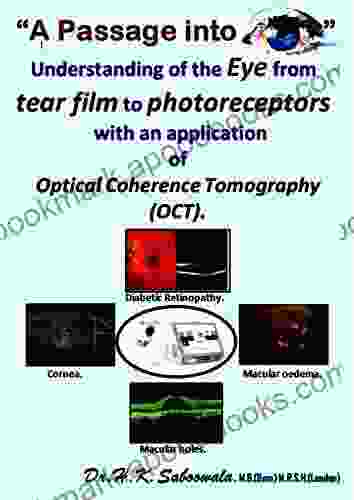
Unveiling the Eye's Intricacies: From Tear Film to Photoreceptors

5 out of 5
| Language | : | English |
| Text-to-Speech | : | Enabled |
| Enhanced typesetting | : | Enabled |
| Lending | : | Enabled |
| File size | : | 11054 KB |
| Screen Reader | : | Supported |
| Print length | : | 39 pages |
The human eye is a remarkable organ that allows us to perceive the world around us. Its intricate structure and complex function have fascinated scientists and medical professionals for centuries. In this article, we will embark on a journey to understand the eye, from the tear film that protects its surface to the photoreceptors that capture light and convert it into electrical signals. Along the way, we'll uncover the intricacies of the cornea, lens, retina, and other vital components that work together to give us the gift of sight.
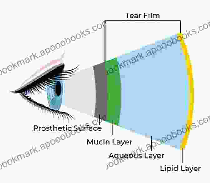
The Tear Film: A Protective Barrier
The tear film is a thin layer of fluid that covers the surface of the eye. It plays a crucial role in protecting the eye from infection, dehydration, and mechanical damage. The tear film is composed of three layers:
- The outer lipid layer is produced by the meibomian glands in the eyelids and helps to prevent evaporation of the tear film.
- The middle aqueous layer is produced by the lacrimal glands and contains water, electrolytes, and proteins that nourish and protect the cornea.
- The inner mucin layer is produced by the goblet cells in the conjunctiva and helps to keep the tear film attached to the surface of the eye.
The tear film is constantly being produced and drained away. This process helps to remove debris and keep the eye surface clean.
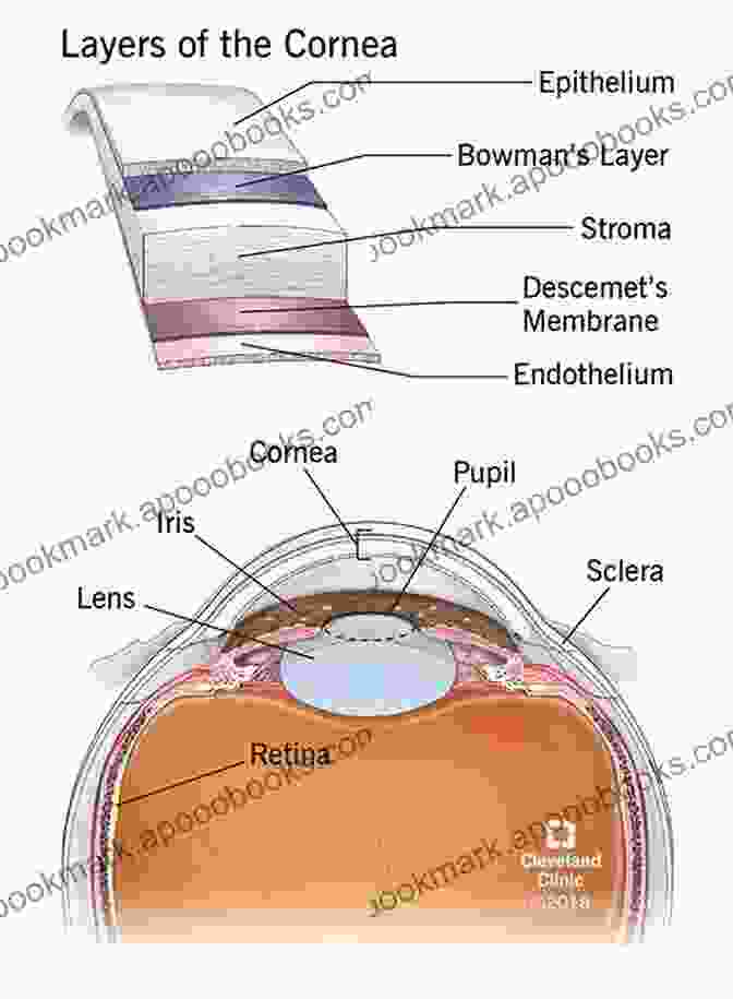
The Cornea: Transparent Window to the World
The cornea is the clear, dome-shaped structure that forms the front of the eye. It is responsible for focusing light on the retina, the light-sensitive tissue at the back of the eye. The cornea is composed of five layers:
- Epithelium: The outermost layer of the cornea is made up of several layers of epithelial cells. These cells help to protect the cornea from infection and mechanical damage.
- Bowman's layer: A thin layer of collagen fibers that provides additional strength to the cornea.
- Stroma: The thickest layer of the cornea, composed of collagen fibers arranged in a regular pattern. The stroma gives the cornea its strength and transparency.
- Descemet's membrane: A thin layer of collagen fibers that lies beneath the stroma.
- Endothelium: The innermost layer of the cornea is made up of a single layer of endothelial cells. These cells help to pump fluid out of the cornea, keeping it clear and transparent.
The cornea is avascular, meaning it does not contain any blood vessels. This allows the cornea to remain clear and transparent, allowing light to pass through it without distortion.
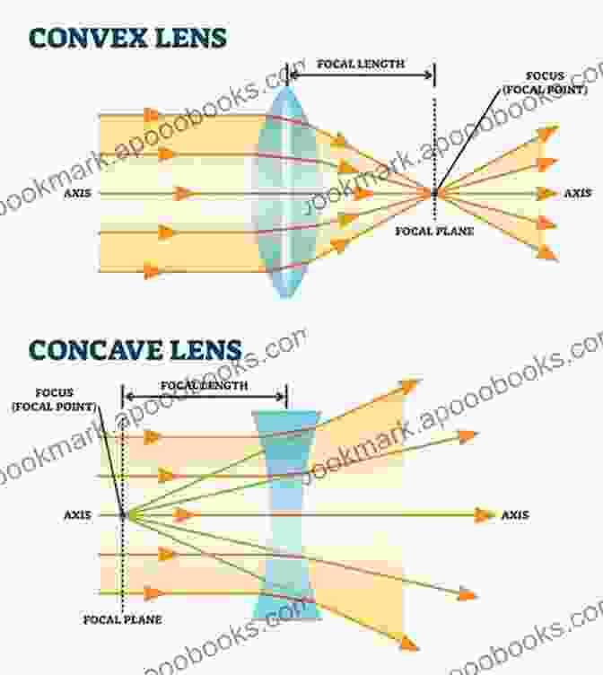
The Lens: Adjustable Focus
The lens is a transparent, biconvex structure that sits behind the cornea. It is responsible for fine-tuning the focus of light on the retina. The lens is composed of three main parts:
- Capsule: A thin membrane that surrounds the lens.
- Cortex: The outer layer of the lens, composed of lens fibers.
- Nucleus: The central core of the lens, composed of older lens fibers.
The lens is able to change its shape to adjust the focus of light on the retina. This process is called accommodation. Accommodation allows us to focus on objects at different distances.
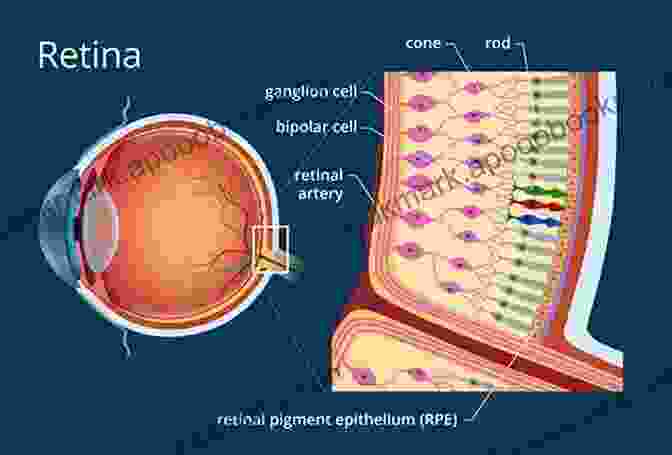
The Retina: Capturing Light
The retina is a thin, light-sensitive tissue that lines the back of the eye. It contains millions of photoreceptors, which are specialized cells that convert light into electrical signals. The retina is divided into two main regions:
- The central retina, which contains the macula, a small area that is responsible for central vision and detailed perception.
- The peripheral retina, which is responsible for peripheral vision and motion detection.
The photoreceptors in the retina are of two types: cones and rods. Cones are responsible for color vision and are most active in bright light conditions. Rods are responsible for black-and-white vision and are more active in low light conditions.
Other Important Structures
In addition to the tear film, cornea, lens, and retina, the eye contains several other important structures, including:
- The iris is the colored part of the eye. It contains muscles that control the size of the pupil, which is the black hole in the center of the iris.
- The pupil is the opening in the iris that allows light to enter the eye.
- The ciliary body is a ring of tissue that surrounds the lens. It contains muscles that help to ac
5 out of 5
| Language | : | English |
| Text-to-Speech | : | Enabled |
| Enhanced typesetting | : | Enabled |
| Lending | : | Enabled |
| File size | : | 11054 KB |
| Screen Reader | : | Supported |
| Print length | : | 39 pages |
Do you want to contribute by writing guest posts on this blog?
Please contact us and send us a resume of previous articles that you have written.
 Book
Book Novel
Novel Page
Page Chapter
 Text
Text Story
Story Genre
Genre Reader
 Library
 Paperback
 E-book
 Magazine
 Newspaper
 Paragraph
 Sentence
 Bookmark
 Shelf
Shelf Glossary
 Bibliography
 Foreword
 Preface
 Synopsis
 Annotation
 Footnote
 Manuscript
 Scroll
 Codex
Codex Tome
Tome Bestseller
 Classics
 Library card
 Narrative
 Biography
 Autobiography
 Memoir
 Reference
 Encyclopedia
 Emma Mieko Candon
 Tom Leddy
 Heather Walpole
 Margot Starbuck
 E Wayne Ross
 Paul Irwing
 Edgar Wallace
 Kristen Ethridge
 Educational Partners International Llc
 Mark Hatala
 Vincent Hunanyan
 Fred Colby
 Meg Bateman
 Sheila Riley
 Durga Chew Bose
 Hadley Wickham
 Michael R Gordon
 P K Mallick
 Joe Wysong
 Elithe Hamilton Kirkland
Light bulbAdvertise smarter! Our strategic ad space ensures maximum exposure. Reserve your spot today!

 Calvin FisherVariants And Other Difficult Diagnoses: A Guide to Unlocking the Secrets of...


 Craig CarterAn Illustrated Guide to How Mistruths Are Sold, Why They Stick, and How to...
 Guillermo BlairUnlock the Secrets of the Classified Report: An Abbreviated Glimpse into the...
 Herb SimmonsFollow ·8.7k
 Nathan ReedFollow ·4k
 Jeffrey HayesFollow ·5k
 Isaac AsimovFollow ·2.2k
 Duane KellyFollow ·16k
 Ernest PowellFollow ·8.5k
 Miguel de CervantesFollow ·6.6k
 Gabriel Garcia MarquezFollow ·12.2k

 Eugene Powell
Fat Cat Stories: Level At Word Family - A Purrfect Start...
Introducing the 'At'...

 William Powell
Unveiling the Treasures of Russian Poetry: The Cambridge...
Immerse yourself in the...

 Roberto Bolaño
Unveiling the Treasures of Beowulf: A Guided Tour with...
: Delving into the...

 Foster Hayes
Transport, Climate Change and the City: Tackling Urban...
Transport is a major...
 Calvin Fisher
How To Make It In The Music Industry: The Ultimate Guide...
Are you an aspiring musician with...

 Rick Nelson
Unveiling the Enigmatic World of Gary Chester's "The New...
Step into a World...
5 out of 5
| Language | : | English |
| Text-to-Speech | : | Enabled |
| Enhanced typesetting | : | Enabled |
| Lending | : | Enabled |
| File size | : | 11054 KB |
| Screen Reader | : | Supported |
| Print length | : | 39 pages |
